Pregnancy and breast feeding cause increases in calcium demand in the body and the body may respond by “mining” the bones for that mineral. The fetal skeleton requires calcium and phosphorus during rapid growth in the final 3 months of pregnancy. Bone weakening can result. Most women do not have problems in the short run. Bone mass reductions during pregnancy and breastfeeding reverse within six months after the birth or cessation of breastfeeding.
However, the temporary decline in bone density may have consequences that show up decades later - it is not clear whether having gone through a pregnancy makes a woman more or less at risk for osteoporosis. When bone loss does occur, it usually returns to normal within a few months of the birth or the end of breastfeeding.
Estrogen levels and swings in the body affect osteoporosis risk, although the mechanics of how this works are not entirely understood. Pregnancy produces enormous effects on steroid hormone levels and longtitudinal studies have shown a woman’s pregnancy history affects osteoporosis risk. Breast feeding, which takes calcium out of the mother’s body, also affects bone density.
Most women do not have significant bone problems during pregnancy and lactation. Modern prenatal care regimens take measures to avoid problems and calcium supplements can help. Further, it has been shown that the pregnant woman’s body absorbs dietary calcium from regular food better than other women’s bodies. This is particularly true in the second half of the pregnancy.
Animal tests suggest that vitamin D deficiency during pregnancy can result in bone problems in the children. Even studies of humans have suggested that bone growth in fetuses and infants can affect osteoporosis risk decades later. The effect of a woman’s pregnancy history on breast cancer risk is often mentioned in discussions of women’s health. Early pregnancies, late pregnancies, not ever getting pregnant or delivering a baby – these are all said to affect chances of developing breast cancer because they affect hormone levels.
One radical step to avoiding breast cancer is called preventive bilateral oophorectomy. It is surgical removal of the ovaries and the idea is sometimes floated for women who have abnormal BRCA1 or BRCA2 genes. (How often this procedure is done is another question.) There are downsides to oophorectomies, including early onset of menopause. No ovaries means reduced levels of estrogen in the body and that can result in lower bone density. A survey found women who had the procedure had over 70% increase in incidence of both osteoporosis and arthritis. Environmental and even emotional stresses can affect the menstrual cycle, and indirectly result in changes in bone density. The effect over a month or two is very small, but long-term modification of estrogen levels makes a difference in osteoporosis risk.
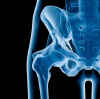
Transient osteoporosis of the hip suddenly occurs sometimes in the third trimester of otherwise healthy pregnancies. It can be immobilizing and painful, but it generally passes within a few weeks after birth. As with transient osteoporosis of the hip in men, the cause is unknown. There have also been reports of an even rarer transient osteoporosis of the knee during pregnancy.
Pregnancy is good for your bones in the long run! You might lose bone mass in the third trimester and breastfeeding, but it comes back stronger. The bone mass returns to normal six months after breastfeeding ends, and there is evidence that the more times a woman has been pregnant (into the third trimester), the lower the risk of fractures later in life.
Lactation and breast feeding, which occurs directly after pregnancy, is also a time of increased incidence of bone weakening. It is easy to understand at least part of the reason why. If the growth of the fetus includes the growth of bone and use of calcium from the mother’s body, the production and removal of milk likewise involves calcium removal from the mother’s body.
The effect of pregnancy and child-birth on future risk for osteoporosis and osteopenia argues for a lifetime assessment and records-keeping system. Perhaps with society-wide adoption of more advanced information management systems our medical system of the future will be equipped with a more complete picture of a patient’s history.
Endurance athletes in training sometimes find their menstrual cycle stops or becomes irregular - oligomenorrheic or amenorrheic. It has been shown that such athletes have higher rates of osteopenia than normal. This applies to things like running. Sports training with resistance tends to increase bone density. A regimen that consists of both – such as cross training – can even result in both at the same time. The small bones not involved in resistance can suffer declines in density due to a drop in estrogen while the larger bones getting the strength training can increase in density.
Stress fractures in bones are common in endurance athletes of both sexes. Scientists have established that non-menstruating or irregular-menstruating women athletes have 2 to 4 times the risk for stress fracture. http://web.stanford.edu/~kcobb/bfit/book/ The hyposthesis is that the low estrogen levels increase the fracture rate, an idea supported by evidence that oral contraceptive use reduces incidence of fractures.